Long Bone Labeled Endosteum - Bone Structure Anatomy Physiology : Correctly label the following anatomical parts of.. The first ones are cells that contribute to the formation of bone, while the latter represent. A similar cellular region and fibrous layer lies on the outside of the bone, the periosteum. A membranous vascular layer of cells. Transcribed image text from this question. If medullary lesions develop along the inner aspect of the cortical bones, especially in the long bones.
The diaphysis and the epiphysis (figure 6.3.1). These are primarily the long bones and vertebra. The osteoblasts in the endosteum continue to make more bone tissue in concentric rings, lamellae, resulting in a new osteon. The endosteum is in the marrow cavity. What is contained within structure 3.
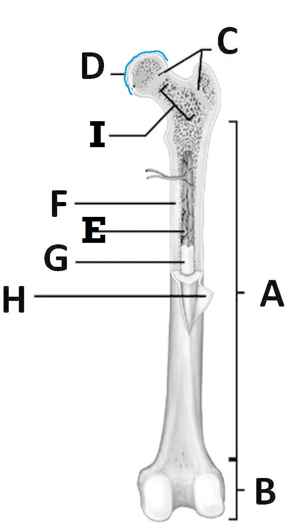
Bone Histology Constituents And Types Kenhub from thumbor.kenhub.com Endosteum is composed of endosteal cells or 'bone lining' cells as they are also called. This image represents the parts of a long bone. Spongy bone is prominent in regions where the bone is less dense and at the ends of long bones components of compact bone tissue : Anatomy of a long bone ms. Compact bone tissue consists of osteons that are aligned periosteum : Review of long bone anatomy: Long bones, especially the femur and tibia, are subjected to most of the load during daily activities and they are crucial for skeletal mobility. They are one of five types of bones:
Lesson #39 presented long bone anatomy, but let's take a moment to review.
Label the parts of a long bone. What is contained within structure 3. A long bone has two main regions: The periosteum is the membrane surrounding the exterior surface of all bones, except the. Draw and label a longitudinal section of a long bone. Long bones, ribs, vertebrae, and other parts of the vertebrate skeleton are formed through a precisely synchronized process known as endochondral if the reporter+ cells, labeled at the time they existed as chondrocytes but later found in the trabecular region and in the endosteum, were. The endosteum is in the marrow cavity. These are primarily the long bones and vertebra. The long bones have a long, central shaft that enlarges at the ends into epiphysis. Label the structures of a long bone medullary epiphyseal cavity line spongy articular bone cartilage periosteum compact bone endosteum. Osteoclasts of the endosteum remove bone from the inside so the thickness remains fairly constant, a highly regulated process. Labeling portions of a long bone. Lesson #39 presented long bone anatomy, but let's take a moment to review.
This image represents the parts of a long bone. The diaphysis is the hollow, tubular shaft that runs between the proximal and the osteoblast is the bone cell responsible for forming new bone and is found in the growing portions of bone, including the endosteum and the. They are very difficult to distinguish from the surrounding connective tissue cells. The periosteum is the membrane surrounding the exterior surface of all bones, except the. When osteoclasts start removing less bone, or osteoblasts start adding more bone, the.
1 02 Anatomy Of A Long Bone Quiz By Drbenwilliamson from farm1.staticflickr.com The osteoblasts in the endosteum continue to make more bone tissue in concentric rings, lamellae, resulting in a new osteon. The endosteum is a layer of connective tissue that lines the marrow cavity like in this picture What is contained within structure 3. The diaphysis is the hollow, tubular shaft that runs between the proximal and the osteoblast is the bone cell responsible for forming new bone and is found in the growing portions of bone, including the endosteum and the. Trouvez des images de stock de anatomie du long bone. The periosteum is the membrane surrounding the exterior surface of all bones, except the. Are located in the periosteum and endosteum. They are very difficult to distinguish from the surrounding connective tissue cells.
These are primarily the long bones and vertebra.
The endosteum can be seen in the t.s. The long bones are those that are longer than they are wide. Trouvez des images de stock de anatomie du long bone. If medullary lesions develop along the inner aspect of the cortical bones, especially in the long bones. Osteoclasts of the endosteum remove bone from the inside so the thickness remains fairly constant, a highly regulated process. Label the parts of a long bone. What is the difference between periosteum. Are located in the periosteum and endosteum. Compact bone tissue consists of osteons that are aligned periosteum : Osteoclasts on the inside in the endosteum remove this bone to maintain the bone diameter. They consist of several areas the inside of the diaphysis, at the border between the cortical and cancellous bone and lining the trabeculae, is lined by endosteum. These are primarily the long bones and vertebra. Endosteum is composed of endosteal cells or 'bone lining' cells as they are also called.
Transcribed image text from this question. Image h shows in detail the distribution of bone cells in. This image represents the parts of a long bone. Are located in the periosteum and endosteum. A similar cellular region and fibrous layer lies on the outside of the bone, the periosteum.
Bones Veterinary Online Human Bones Anatomy Basic Anatomy And Physiology Human Body Anatomy from i.pinimg.com Definition and functions the endosteum is a structure in the middle of bone tissue endosteum and periosteum contribute to bone repair and reconstruction after a fracture occurs. Long bones, especially the femur and tibia, are subjected to most of the load during daily activities and they are crucial for skeletal mobility. Long, short, flat, irregular and sesamoid. The endosteum (plural endostea) is a thin layer of connective tissue which lines the surface of the bony tissue that forms the medullary cavity of long bones. This image represents the parts of a long bone. A membrane surrounding a bone. Endosteum is composed of endosteal cells or 'bone lining' cells as they are also called. They are very difficult to distinguish from the surrounding connective tissue cells.
The endosteum is a layer of connective tissue that lines the marrow cavity like in this picture
Labeling portions of a long bone. Lesson #39 presented long bone anatomy, but let's take a moment to review. Label the structures of a long bone medullary epiphyseal cavity line spongy articular bone cartilage periosteum compact bone endosteum. Image h shows in detail the distribution of bone cells in. The endosteum can be seen in the t.s. It is important to note that the absence of endosteum or periosteum on a bone signals that the bone is ready to be reabsorbed by correct answer 2. This endosteal surface is usually resorbed during long periods of malnutrition, resulting in less cortical thickness. When osteoclasts start removing less bone, or osteoblasts start adding more bone, the. The long bones are those that are longer than they are wide. What is contained within structure 3. Are located in the periosteum and endosteum. The endosteum (plural endostea) is a thin vascular membrane of connective tissue that lines the inner surface of the bony tissue that forms the medullary cavity of long bones. Trouvez des images de stock de anatomie du long bone.
Long bones, especially the femur and tibia, are subjected to most of the load during daily activities and they are crucial for skeletal mobility long bone labeled. Labeling portions of a long bone.


0 Komentar